Table of Contents >> Show >> Hide
- What Is a Mammary Duct?
- Basic Breast Anatomy: Where the Mammary Ducts Fit
- Mammary Duct Diagram
- How Mammary Ducts Work
- Terminal Duct Lobular Unit: The Functional Center
- Mammary Ducts During Puberty, Pregnancy, and Breastfeeding
- Common Conditions Involving Mammary Ducts
- Symptoms That May Involve the Milk Ducts
- How Doctors Examine the Mammary Duct System
- Why Mammary Duct Anatomy Matters
- Experience-Based Insights: Understanding Mammary Ducts in Everyday Life
- Conclusion
Note: This article is for educational purposes only and is not a substitute for professional medical advice, diagnosis, or treatment.
The mammary duct may sound like a tiny backstage hallway in the body, but it plays a starring role in breast anatomy, breastfeeding, and breast health. These small tubes are part of the ductal system of the breast, carrying milk from milk-producing glands to the nipple. Simple job? On paper, yes. In real life, the mammary duct system is a beautifully organized network of lobes, lobules, ducts, nerves, blood vessels, lymph channels, fat, and connective tissue all working together like a very committed production crew.
Understanding mammary duct anatomy helps explain how lactation works, why breast changes can happen during puberty, pregnancy, breastfeeding, menstruation, and menopause, and why doctors pay close attention to the ducts during breast imaging. Many breast conditions, including duct ectasia, plugged ducts, mastitis, ductal carcinoma in situ, and invasive ductal carcinoma, involve the milk ducts. That does not mean every duct-related symptom is dangerous, but it does mean the duct system deserves more attention than it usually gets.
This guide breaks down the anatomy, function, and diagram of the mammary ducts in clear, friendly American Englishno medical-school decoder ring required.
What Is a Mammary Duct?
A mammary duct, also called a milk duct or lactiferous duct, is a small tube inside the breast that transports milk from the lobules to the nipple during breastfeeding. Mammary ducts are part of the glandular tissue of the breast. They are most developed in people assigned female at birth after puberty, but all sexes have some breast duct tissue beneath the nipple.
The word “mammary” refers to the breast or milk-producing glands. The duct itself does not make milk. Instead, it works more like a delivery route. Milk is produced in tiny glandular structures called lobules and alveoli, then moves through smaller ducts that merge into larger ducts near the nipple.
Think of the system like a tree: the lobules are the leaves and fruit, the small ducts are twigs, the larger ducts are branches, and the nipple is where the whole tree politely hands over the milk. Biology may not always be poetic, but in this case, it is surprisingly well-plumbed.
Basic Breast Anatomy: Where the Mammary Ducts Fit
The breast is made of several types of tissue. The main components include glandular tissue, fatty tissue, connective tissue, blood vessels, lymph vessels, nerves, the nipple, and the areola. Mammary ducts sit within the glandular portion of the breast, connecting milk-producing structures to the nipple surface.
Lobes
Each breast contains multiple sections called lobes. Many anatomy references describe the breast as having about 15 to 20 lobes, although the exact number can vary from person to person. These lobes are arranged somewhat like petals around the nipple.
Lobules
Inside each lobe are smaller structures called lobules. Lobules are the milk-producing glands of the breast. During pregnancy and breastfeeding, hormonal signals prepare these lobules to produce milk.
Alveoli
Within the lobules are tiny sac-like units called alveoli. These are lined with milk-secreting cells. When lactation is active, alveoli produce milk and release it into nearby small ducts.
Mammary Ducts
The mammary ducts form the transport network. Small ducts collect milk from the alveoli and lobules. These small ducts join into larger ducts, which move toward the nipple. Near the nipple, several ducts open onto the surface through tiny nipple pores.
Nipple and Areola
The nipple is the raised central structure where milk exits the breast. The areola is the darker circular area around the nipple. The areola contains small glands that help lubricate and protect the skin, especially during breastfeeding.
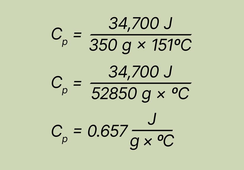
Mammary Duct Diagram
The following simple diagram shows how the mammary duct system connects the milk-producing parts of the breast to the nipple:
How Mammary Ducts Work
The main function of mammary ducts is to carry milk. During lactation, milk production and release depend on a combination of glandular tissue, hormones, nerves, and muscle-like cells around the milk-producing units.
Step 1: Hormones Prepare the Breast
During pregnancy, hormones such as estrogen, progesterone, prolactin, and others help prepare the breast for lactation. Lobules and ducts become more developed. Many people notice breast fullness, tenderness, darker areolas, and more visible veins during this time.
Step 2: Lobules Produce Milk
After childbirth, prolactin helps signal the alveoli in the lobules to make milk. Milk production is not happening randomly; it is controlled by a supply-and-demand system. Frequent milk removal through breastfeeding or pumping tells the body to continue producing milk.
Step 3: Milk Moves Through the Ducts
When a baby suckles, nerve signals travel from the nipple and areola to the brain. The hormone oxytocin is released, causing tiny muscle-like myoepithelial cells around the alveoli and ducts to contract. This pushes milk through the mammary ducts toward the nipple. This process is called the let-down reflex.
Step 4: Milk Leaves Through the Nipple
Milk exits through small openings on the nipple surface. Not every duct has a large visible opening, and the number of duct openings varies. The system is efficient, compact, and much more sophisticated than it looks from the outside.
Terminal Duct Lobular Unit: The Functional Center
One important structure in breast anatomy is the terminal duct lobular unit, often shortened to TDLU. This unit includes a lobule and its small associated duct. It is considered the functional unit of the breast because it is where milk production and early duct-lobule activity occur.
The TDLU is also medically important because many breast cancers and benign breast conditions begin in or near the ductal and lobular structures. This is one reason mammograms, ultrasounds, MRIs, and biopsies often focus on changes within ductal or glandular tissue.
Mammary Ducts During Puberty, Pregnancy, and Breastfeeding
During Puberty
At puberty, estrogen and other hormones stimulate breast development. Ducts grow and branch into the surrounding fatty tissue. The amount of fat, connective tissue, and glandular tissue determines breast size and density. Larger breasts do not necessarily mean more ducts or better milk production. Breast size is mostly influenced by fatty tissue, not duct performance.
During Pregnancy
Pregnancy causes major changes in the duct and lobule system. Lobules enlarge, ducts become more active, and the breast prepares for milk production. The areola may darken, the nipple may become more prominent, and the breast may feel heavier or more sensitive.
During Breastfeeding
During breastfeeding, mammary ducts transport milk repeatedly throughout the day. Milk flow may feel like tingling, tightening, warmth, or a sudden sense of fullness during let-down. Some people feel the let-down reflex clearly; others barely notice it. Both experiences can be normal.
After Weaning
After breastfeeding ends, milk production gradually decreases. The ducts and lobules become less active, and the breast tissue changes again. Some fullness or occasional leakage can happen for a while, but persistent or unusual nipple discharge should be discussed with a healthcare professional.
Common Conditions Involving Mammary Ducts
Because mammary ducts are tiny tubes, they can become inflamed, blocked, widened, or affected by abnormal cell growth. Many duct-related conditions are benign, but new or persistent symptoms should not be ignored.
Plugged Milk Duct
A plugged duct can happen during breastfeeding when milk flow is reduced in one area. It may feel like a tender lump, firm spot, or localized area of swelling. Modern breastfeeding guidance often focuses on reducing inflammation, avoiding aggressive massage, feeding or pumping normally, and contacting a healthcare provider if symptoms worsen or fever develops.
Mastitis
Mastitis is inflammation of breast tissue that may involve infection. It can cause breast pain, swelling, warmth, redness, fever, chills, and flu-like symptoms. Mastitis can happen during breastfeeding but may also occur in other situations. Prompt care is important because untreated infection can sometimes lead to an abscess.
Mammary Duct Ectasia
Mammary duct ectasia is a noncancerous condition in which one or more milk ducts beneath the nipple widen, thicken, and may fill with fluid. It is more common around perimenopause and menopause. Some people have no symptoms, while others may notice nipple tenderness, sticky discharge, nipple inversion, or a lump behind the nipple. Although it is benign, it can mimic more concerning conditions, so evaluation is often recommended.
Intraductal Papilloma
An intraductal papilloma is a small benign growth inside a milk duct. It often appears near the nipple and may cause clear or bloody nipple discharge. While usually noncancerous, it should be checked because symptoms can overlap with other breast conditions.
Ductal Carcinoma in Situ
Ductal carcinoma in situ, or DCIS, involves abnormal cells contained within a milk duct. It is considered noninvasive because the abnormal cells have not spread into surrounding breast tissue. DCIS is often found on mammograms because of tiny calcium deposits called calcifications.
Invasive Ductal Carcinoma
Invasive ductal carcinoma begins in a milk duct and then grows into nearby breast tissue. It is the most common type of breast cancer. This is why understanding mammary duct anatomy is not just interestingit is clinically important.
Symptoms That May Involve the Milk Ducts
Duct-related symptoms can be harmless, hormonal, infectious, or more serious. A healthcare provider can help determine the cause. Symptoms that deserve medical attention include:
- A new breast lump or thickened area
- Bloody, clear, green, yellow, or persistent nipple discharge not related to breastfeeding
- A nipple that suddenly turns inward
- Skin dimpling, puckering, scaling, or redness
- Breast pain that does not improve
- Warmth, swelling, fever, or flu-like symptoms
- Changes in breast shape or nipple appearance
Many breast changes are not cancer, but checking them is the wise move. Breasts are not the place to practice heroic denial. If something is new, unusual, or persistent, get it evaluated.
How Doctors Examine the Mammary Duct System
Clinical Breast Exam
A clinician may examine the breast, nipple, areola, and underarm area to check for lumps, tenderness, discharge, swelling, or lymph node changes.
Mammogram
A mammogram is an X-ray of the breast. It can detect calcifications, masses, and architectural changes that may involve ducts or lobules. Mammograms are especially important for breast cancer screening.
Ultrasound
Breast ultrasound uses sound waves to evaluate lumps, fluid-filled cysts, duct changes, abscesses, and areas that need closer inspection. It is often used along with mammography.
Breast MRI
Breast MRI may be used for high-risk screening, evaluating the extent of certain cancers, or examining complex breast findings. It provides detailed images of soft tissue and blood flow patterns.
Biopsy
If imaging shows a suspicious area, a biopsy may be performed. A small tissue sample is removed and examined under a microscope. This is the only way to confirm whether abnormal cells are benign, precancerous, or cancerous.
Why Mammary Duct Anatomy Matters
Mammary ducts matter because they connect anatomy, function, and health. They are essential for breastfeeding, but they are also involved in many common breast symptoms and diagnoses. Knowing the basic structure of the duct system can make medical conversations easier and less intimidating.
For example, when a doctor says a finding is “ductal,” it usually means it begins in or relates to the milk ducts. When a lactation consultant talks about milk transfer, let-down, or plugged ducts, they are talking about how well milk moves through this duct network. When a radiology report mentions duct dilation, calcifications, or a retroareolar finding, it often points to the area beneath or near the nipple where ducts converge.
In other words, mammary ducts may be small, but they show up in big conversations: breastfeeding, breast pain, nipple discharge, cancer screening, and breast surgery.
Experience-Based Insights: Understanding Mammary Ducts in Everyday Life
For many people, mammary duct anatomy becomes real only when something changes. Before that, the breast may feel like one general structure: soft tissue, nipple, areola, done. Then pregnancy, breastfeeding, a mammogram, or a sudden symptom turns the duct system from “anatomy trivia” into something practical and personal.
One common experience is the surprise of let-down during breastfeeding. A parent may expect milk to simply appear, but the process can feel dramatic: tingling, pressure, leaking from the opposite breast, or a sudden change in the baby’s swallowing rhythm. That moment is mammary duct function in action. The lobules produce milk, oxytocin helps push it forward, and the ducts carry it to the nipple. The body does not ask for applause, but honestly, it earns some.
Another relatable experience is dealing with a tender spot during breastfeeding. A person may feel a firm area and immediately worry. Sometimes this is related to inflammation and reduced milk movement in a section of the duct system. The key lesson is that ducts are delicate. They are not garden hoses that need forceful squeezing. Gentle care, normal milk removal, supportive guidance, and medical help when symptoms worsen are far better than aggressive pressure or panic-Googling at 2 a.m.
People who are not breastfeeding can also become aware of their ducts through nipple discharge or changes behind the nipple. A small amount of discharge may have many causes, but persistent, bloody, spontaneous, or one-sided discharge deserves evaluation. This is where understanding duct anatomy reduces fear while encouraging action. The symptom may be benign, such as duct ectasia or a papilloma, but a clinician can determine what is happening.
Breast imaging is another moment when duct anatomy becomes useful. A mammogram report may mention calcifications, density, asymmetry, or ductal changes. These words can sound alarming, especially when they appear in a patient portal before a doctor explains them. Knowing that ducts and lobules are normal breast structures can make the language less frightening. It also helps people ask better questions, such as: “Is this finding in a duct?” “Do I need ultrasound?” “Is biopsy recommended?” “How soon should I follow up?”
From an educational perspective, the best way to visualize mammary ducts is to imagine grapes on stems. The grapes are the lobules and alveoli, the stems are the ducts, and the nipple is the exit point. This simple picture is not perfect, but it helps. It explains why a problem in one small area can create a localized lump or tender spot, and why breast tissue can feel different depending on hormones, age, lactation, and body composition.
The most important experience-based takeaway is this: breast awareness is not about obsessively checking every tiny sensation. It is about knowing your own normal. Breasts naturally change during menstrual cycles, pregnancy, breastfeeding, weight changes, and aging. But new, persistent, or unusual changes should be checked. A calm appointment beats weeks of worry, and early evaluation is always smarter than guessing.
Conclusion
The mammary duct is a small but essential part of breast anatomy. It carries milk from the lobules to the nipple, supports breastfeeding, and plays a major role in many breast health conditions. The duct system begins in tiny milk-producing structures, branches through the breast, and converges near the nipple like a carefully designed biological delivery network.
Understanding mammary duct anatomy, function, and diagram basics can help make sense of lactation, plugged ducts, nipple discharge, breast imaging, and duct-related diagnoses. While many duct changes are benign, symptoms such as persistent discharge, new lumps, nipple inversion, fever, or skin changes should be evaluated by a healthcare professional.
In short, mammary ducts may be tiny tubes, but they carry a big responsibility. They help nourish infants, explain many breast changes, and provide important clues in breast health. Small structure, major résumé.