Table of Contents >> Show >> Hide
- Inner ear basics: where it is and what it does
- An anatomy tour of the inner ear (no lab coat required)
- How hearing works (in plain English)
- How balance works (and why your brain sometimes freaks out)
- Common inner ear conditions (what they are and how they typically feel)
- 1) Benign paroxysmal positional vertigo (BPPV)
- 2) Vestibular neuritis and labyrinthitis
- 3) Ménière’s disease
- 4) Vestibular migraine (a frequent “surprise” cause of vertigo)
- 5) Tinnitus and noise-induced inner ear damage
- 6) Sudden sensorineural hearing loss (SSHL): treat it like an emergency
- 7) Acoustic neuroma (vestibular schwannoma)
- 8) Otosclerosis (not inner ear, but it can affect inner ear function)
- 9) Superior canal dehiscence syndrome (SCDS)
- 10) Perilymphatic fistula (PLF)
- 11) Autoimmune inner ear disease (AIED)
- How inner ear problems are diagnosed
- Treatment and self-care: what usually helps (and when to seek care fast)
- How to protect your inner ear (future-you will thank you)
- Quick FAQs
- Conclusion
- Real-world experiences with inner ear issues (about )
- SEO tags (JSON)
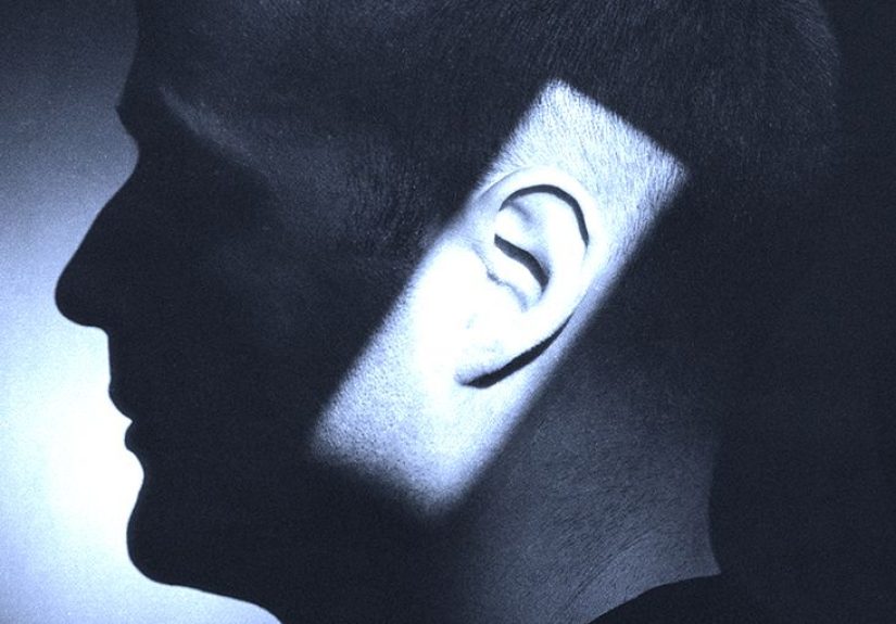
Your inner ear is basically a tiny, bone-protected “theme park” that runs two of your biggest life upgrades:
hearing and balance. It’s smaller than a grape, hidden deep in your skull,
and yet it can make you enjoy music, understand speech, stay upright, and walk in a straight line without looking like you’re auditioning for a slapstick comedy.
When it’s happy, you never think about it. When it’s mad? Suddenly the room is spinning, your ears are ringing, and you’re Googling “Why does my head hate me?”
This guide breaks down inner ear anatomy, how it works, and the most common inner ear conditionsplus what doctors typically look for and
what you can do to protect this delicate (and slightly dramatic) system.
Inner ear basics: where it is and what it does
The inner ear sits inside the temporal bone (the thick bone on the side of your skull). Think of it as a paired set of systems:
- The cochlea: your hearing “translator,” turning sound vibrations into nerve signals.
- The vestibular system: your balance “navigation team,” tracking head movement and orientation.
Both systems connect to your brain through the vestibulocochlear nerve (cranial nerve VIII), which splits into hearing and balance branches.
An anatomy tour of the inner ear (no lab coat required)
The cochlea: a snail shell that hears
The cochlea is a spiral-shaped, fluid-filled structure. When sound energy reaches it, waves travel through its fluids and move a specialized
membrane where sensory hair cells live (yes, real cells with tiny hair-like projections). These hair cells bend in response to vibration and convert motion into
electrical signalsyour brain’s preferred language.
A neat trick: the cochlea is organized by pitch. Different areas respond best to different frequencies (high-pitched sounds vs. low-pitched sounds),
like a built-in sound map. That’s part of why damage can affect some frequencies more than others.
The vestibule: the “gravity and straight-line motion” department
The vestibule contains the otolith organs: the utricle and saccule. These help you sense
linear acceleration (moving forward/backward or up/down) and your head’s position relative to gravity.
Otolith organs rely on tiny calcium carbonate crystals called otoconia. They add weight so movement can tug on hair cellslike a snow globe that shifts when you tilt it.
Most days, this is great. On a bad day, those crystals can wander into places they don’t belong (we’ll get to BPPV).
The semicircular canals: your rotation sensors
You have three semicircular canals in each ear, arranged roughly at right angleskind of like a 3D gyroscope. They detect
rotational movement (turning your head side to side, nodding, tilting).
Inside each canal is fluid and a flexible structure that moves when your head rotates. That movement bends hair cells and sends rapid signals to your brain.
This system also powers an underrated superpower: the vestibulo-ocular reflex (VOR), which helps your eyes stay focused while your head moves.
(Try reading a street sign while walkingthank your VOR.)
The inner ear’s fluids: tiny plumbing, big consequences
The inner ear uses carefully controlled fluids (commonly discussed as endolymph and perilymph) and delicate membranes.
When pressure or fluid balance is offdue to inflammation, leaks, or “third-window” conditionssymptoms like vertigo, hearing changes, or sound sensitivity can show up.
The wiring: the vestibulocochlear nerve
Signals from the cochlea and vestibular organs travel along cranial nerve VIII into the brainstem, where they’re processed and integrated with vision and body-position signals.
That integration explains why dizziness can feel so weird: you’re not just “off balance”your senses may be sending conflicting updates.
How hearing works (in plain English)
Sound waves enter the outer ear, vibrate the eardrum, and get amplified by the middle ear bones. The last of these bones pushes on the inner ear, creating fluid waves in the cochlea.
Those waves bend hair cells, which trigger nerve signals that your brain interprets as speech, music, or your neighbor’s leaf blower at 7 a.m.
Because cochlear hair cells are delicate, they can be damaged by loud noise, aging, certain medications, and some medical conditions. Unlike many other cells,
human inner-ear hair cells generally don’t regenerate wellso prevention is a big deal.
How balance works (and why your brain sometimes freaks out)
Balance is teamwork. Your brain combines input from:
- Vestibular system (inner ear): motion and orientation
- Vision: what you see
- Proprioception: body position sense (signals from muscles and joints)
When one system sends signals that don’t match the otherslike inner ear trouble, visual motion, dehydration, or migraine-related changesyou can feel dizzy,
unsteady, or nauseated. It’s less “my ears are broken” and more “my brain can’t reconcile the meeting notes.”
Common inner ear conditions (what they are and how they typically feel)
1) Benign paroxysmal positional vertigo (BPPV)
BPPV is one of the most common causes of vertigo. It happens when otoconia (those tiny crystals) break loose and drift into a semicircular canal,
especially the posterior canal. When you move your head, the crystals shift and trigger a strong spinning sensation.
Typical pattern: brief episodes (seconds to under a minute) of intense vertigo triggered by position changesrolling over in bed, looking up, bending down.
What helps: clinicians often use a bedside test (like the Dix–Hallpike maneuver) and treat with canalith repositioning (commonly the Epley maneuver),
which uses guided head movements to relocate the crystals to a less annoying part of the inner ear.
2) Vestibular neuritis and labyrinthitis
These are usually linked to inflammation, often after a viral illness.
Vestibular neuritis affects the vestibular nerve and typically causes prolonged vertigo without major hearing loss.
Labyrinthitis involves the labyrinth and can include vertigo and hearing symptoms.
Typical pattern: sudden severe vertigo lasting hours to days, often with nausea and trouble walking. People often improve gradually, but imbalance can linger.
Because certain strokes can mimic these symptoms, clinicians take new, severe vertigo seriouslyespecially if there are neurologic symptoms (weakness, trouble speaking, severe headache).
What helps: short-term symptom control, hydration, andonce the worst passesvestibular rehabilitation to help the brain recalibrate.
3) Ménière’s disease
Ménière’s disease is associated with episodes of vertigo and inner-ear symptoms believed to involve abnormal fluid dynamics (often described as endolymphatic hydrops).
Many people experience:
- Vertigo attacks lasting minutes to hours
- Fluctuating hearing loss (often low-frequency early on)
- Tinnitus (ringing or roaring)
- Aural fullness (pressure or “stuffed ear” feeling)
What helps: management varies, but commonly includes lifestyle steps (often a lower-sodium diet, moderating caffeine/alcohol, sleep and stress support),
and medications for symptom control. Because the condition can look like other disorders, an ENT/audiology evaluation matters.
4) Vestibular migraine (a frequent “surprise” cause of vertigo)
Not all vertigo comes from the inner ear itself. Vestibular migraine can cause vertigo, motion sensitivity, and imbalance with or without a headache.
Triggers often resemble typical migraine triggerssleep disruption, stress, dehydration, certain foods, or hormonal shifts.
5) Tinnitus and noise-induced inner ear damage
Tinnitus is the perception of sound (ringing, buzzing, hissing) without an external source. It can be linked to inner ear hair-cell damage,
including from loud noise exposure. Sometimes tinnitus has treatable causes (wax, infection, medication side effects), and sometimes it’s part of long-term hearing changes.
What helps: treating the underlying cause when possible, hearing protection, hearing aids when appropriate, and sound therapy/behavioral strategies for distress.
The goal is often “reduce impact,” not “flip an off switch.”
6) Sudden sensorineural hearing loss (SSHL): treat it like an emergency
Sudden sensorineural hearing loss can happen all at once or over a few days, often in one ear.
It’s considered an urgent problem because early treatment can matter.
Watch for: sudden hearing drop, muffled sound, ear fullness, tinnitus, or dizzinessespecially if it’s new and one-sided.
What to do: seek prompt medical evaluation (urgent care/ER/ENT), rather than waiting it out.
7) Acoustic neuroma (vestibular schwannoma)
An acoustic neuroma is a usually slow-growing, benign tumor that develops on the nerve connecting the inner ear to the brain.
It can cause one-sided hearing loss, tinnitus, and balance problems. Evaluation often includes hearing testing and imaging.
Management might involve monitoring, surgery, or focused radiation, depending on size, growth, symptoms, and patient factors.
8) Otosclerosis (not inner ear, but it can affect inner ear function)
Otosclerosis is a bone-remodeling condition that commonly affects the stapes bone in the middle ear, reducing its movement.
That limits how efficiently sound energy reaches the cochlea, causing conductive hearing loss. In some cases, it can also be associated with sensorineural components.
Treatments may include hearing aids or surgery in selected cases.
9) Superior canal dehiscence syndrome (SCDS)
In superior canal dehiscence, there is an opening (“dehiscence”) in bone over part of a semicircular canal.
This can create a “third window” that changes how sound and pressure affect the inner ear.
Possible clues: dizziness triggered by loud sounds or pressure changes, unusual awareness of internal sounds (like your own voice), and sound sensitivity.
Diagnosis typically involves specialized testing and imaging. Treatment depends on severity and may include surgery.
10) Perilymphatic fistula (PLF)
A perilymphatic fistula is an abnormal connection that can allow inner ear fluid to leak toward the middle ear.
It’s often discussed in relation to trauma, barotrauma (pressure injury), or sudden strain.
Possible clues: dizziness/imbalance and hearing changes that worsen with pressure changes (altitude shifts, heavy lifting, coughing/sneezing).
Diagnosis can be tricky because symptoms overlap with other vestibular disorders.
11) Autoimmune inner ear disease (AIED)
AIED is a rare condition where immune-related inflammation affects the inner ear, often causing progressive or fluctuating
sensorineural hearing loss, sometimes with balance symptoms. It tends to require specialist care because treatment may involve steroids or other immune-directed therapy,
and clinicians also look for associated systemic autoimmune disease.
How inner ear problems are diagnosed
Because “dizzy” can mean a hundred different things, diagnosis starts with pattern recognition:
- Timing: seconds vs. minutes vs. hours/days
- Triggers: head position (BPPV) vs. spontaneous attacks (Ménière’s, migraine)
- Hearing symptoms: hearing loss, tinnitus, ear fullness
- Neurologic symptoms: weakness, numbness, trouble speaking, severe headache (red flags)
Common tools include a physical exam focused on eye movements (nystagmus), balance tests, hearing tests (audiogram),
and vestibular testing when needed. Imaging may be used when the story suggests a central cause or certain structural conditions.
Treatment and self-care: what usually helps (and when to seek care fast)
Treatment depends on the causethere isn’t one universal fix. But a few themes come up often:
- Repositioning maneuvers for BPPV (often dramatic, usually effective).
- Vestibular rehabilitation to retrain balance after neuritis/labyrinthitis or persistent dizziness.
- Hearing support (hearing aids, assistive devices, and in specific cases, implants).
- Lifestyle strategies (sleep, hydration, trigger management for vestibular migraine; dietary strategies for Ménière’s in some patients).
Seek urgent evaluation if you notice:
- Sudden hearing loss (especially one-sided), with or without dizziness
- New, severe vertigo plus neurologic symptoms (weakness, facial droop, trouble speaking, severe headache)
- Vertigo after significant head injury, or symptoms that are rapidly worsening
How to protect your inner ear (future-you will thank you)
- Respect loud noise. Use hearing protection at concerts, clubs, power tools, and other high-volume environments.
- Don’t ignore persistent ringing or hearing changes. Early evaluation can rule out treatable causes.
- Review medications with a clinician if you’re concerned about hearing or balance side effects.
- Build fall-proof habits if dizziness is recurring: good lighting, stable footwear, handrails, and avoiding risky ladders when symptomatic.
Quick FAQs
Is dizziness always an inner ear problem?
Nope. Inner ear issues are common causes of true vertigo (spinning sensation), but dizziness can also come from migraine, blood pressure changes,
dehydration, medication effects, anxiety, anemia, heart rhythm issues, and neurological conditions. The detailstiming, triggers, and hearing symptomshelp narrow it down.
Can earwax cause vertigo?
Earwax and middle ear problems more often cause muffled hearing, fullness, or imbalancebut they can contribute to dizziness in some people.
True spinning vertigo is often vestibular (inner ear) or neurologic, so persistent symptoms deserve evaluation.
Why does vertigo make people nauseated?
Your balance system is tightly linked to eye movement and brainstem centers that coordinate nausea and vomiting.
When your brain senses motion that doesn’t match visual or body cues, it can respond like you’ve been poisoned (which is… not the vibe).
Conclusion
The inner ear is small, complex, and ridiculously important. The cochlea turns vibration into hearing, and the vestibular system keeps you oriented in space.
When something goes wronglike displaced crystals (BPPV), inflammation (neuritis/labyrinthitis), fluid dysregulation (Ménière’s), migraine-related changes,
or rare structural/immune issuessymptoms can be dramatic but often treatable.
If there’s one takeaway, it’s this: pattern matters. How long symptoms last, what triggers them, and whether hearing changes are involved can point toward the cause.
And if hearing suddenly drops, don’t waitget checked promptly.
Real-world experiences with inner ear issues (about )
People often describe inner ear symptoms as “impossible to explain until you’ve felt it,” because the experience can be both physical and oddly psychological.
Take the classic BPPV story: someone rolls over in bed, and suddenly the ceiling feels like it’s doing cartwheels. The episode might last only 20 seconds,
but it’s long enough to convince them their brain has entered a new dimension. After a few repeatsturning the head to rinse shampoo, looking up at a shelf,
bending down to tie a shoemany people start moving like a cautious robot, because they’re trying to avoid the “spin switch.” When BPPV is diagnosed,
the treatment can feel like a magic trick: a clinician guides a sequence of head positions, and within minutes the vertigo can dramatically improve.
That sudden relief is why some patients describe the Epley maneuver as “therapy,” “science,” and “a trust fall” all at once.
Vestibular neuritis and labyrinthitis often show up differently. A common experience is waking up after a recent cold and realizing that standing is a bad idea.
The vertigo can be intense enough that the person keeps one hand on the wall, one hand on the bed, and a firm belief that the floor is misbehaving.
Even after the worst passes, the lingering imbalance can be frustrating: walking through a grocery store aisle might trigger symptoms because the visual motion
overwhelms a still-recovering vestibular system. Many people say the turning point comes when they start vestibular rehabilitation exercises.
The exercises can feel counterintuitive“You want me to move my head in the direction that makes me dizzy?”but the logic is retraining the brain’s calibration.
Over time, the brain learns to trust other inputs again, and the world stops feeling like it’s on a slow, rude carousel.
Tinnitus experiences are often more about persistence than intensity. Some people notice ringing after a loud concert and assume it will fade in an hourthen it doesn’t.
Others describe it as a hiss that gets louder at bedtime, when the room is quiet and there’s nothing to mask the sound. The emotional reaction is real:
worry can make tinnitus feel louder, and stress can turn a background noise into a spotlight. People who cope best often build a practical toolkit:
protecting their ears going forward, using a fan or gentle sound at night, and getting hearing checked to see if there’s an underlying hearing loss that can be supported.
For Ménière’s-like symptoms, many people talk about unpredictability as the hardest partnever knowing if an episode will hit during a meeting, a commute, or a family event.
That uncertainty is why structured plans (medical follow-up, trigger tracking, and safety strategies) can be calming even before symptoms fully improve.