Table of Contents >> Show >> Hide
- What Is the Pericardium?
- Pericardium Function: What Does It Actually Do?
- Why the Pericardium Matters in Real Life (Not Just on Exams)
- Common Pericardial Conditions (and What They Look Like)
- How Doctors Evaluate Pericardial Disease
- Treatment Options: From Anti-Inflammatories to Surgery
- When to Get Help
- Conclusion
- Experiences: What People Commonly Go Through With Pericardial Problems (About )
Your heart is a hardworking overachiever. It beats around the clock, never asks for vacation days,
and still manages to look good in every anatomy textbook photo. But even the heart needs a
reliable sidekickand that’s where the pericardium comes in.
Think of the pericardium as your heart’s protective “jacket”: it helps keep the heart in place,
reduces friction as it beats, and can (unfortunately) become the site of problems like
pericarditis, pericardial effusion, and even
cardiac tamponade. In this guide, we’ll break down what the pericardium does,
why it matters, and what happens when it’s not having a great day.
Quick note: This article is for education, not a substitute for personal medical care.
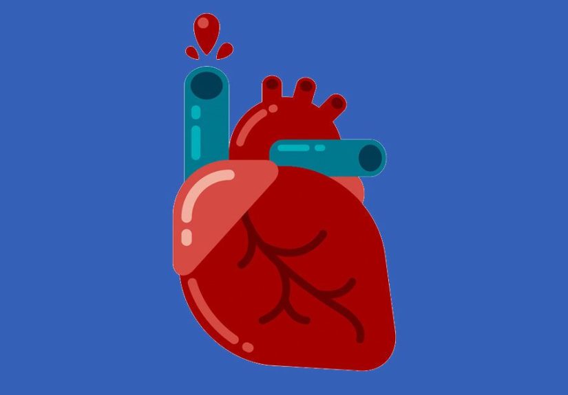
What Is the Pericardium?
The pericardium (also called the pericardial sac) is a thin,
double-layered structure that surrounds your heart and the roots of the big blood vessels leaving and entering it.
It’s not the heart muscle itselfit’s the wrapping around it.
The Two Main Parts: Fibrous and Serous
The pericardium has two major components working like a “tough outer shell + smooth inner lining” combo:
-
Fibrous pericardium: the sturdy outer layer made mostly of connective tissue. It’s the “seatbelt”
that helps prevent your heart from shifting around too much. -
Serous pericardium: the inner, slick layer that reduces friction. It has two sublayers:
- Parietal layer: lines the inside of the fibrous pericardium.
- Visceral layer (also known as the epicardium): hugs the heart’s surface.
The Pericardial Space: A Tiny Slip ’N Slide
Between the parietal and visceral layers is the pericardial cavity, which normally contains a small
amount of lubricating fluid. This fluid lets the heart move smoothly as it contractsbecause a squeaky heart is not
the vibe.
Pericardium Function: What Does It Actually Do?
If the pericardium were a job listing, it would read: “Protect the heart, stabilize it, reduce friction,
and occasionally cause dramatic emergencies.” Here are its greatest hits.
1) Keeps the Heart Positioned and Supported
The pericardium helps anchor the heart within the chest so it doesn’t slosh around like a phone in an unzipped pocket.
This stabilization becomes especially important when you change positions, breathe deeply, or exercise.
2) Reduces Friction During Every Beat
The heart beats tens of thousands of times a day. Without lubrication, the constant motion against nearby tissues
would create friction and irritation. The serous layers plus pericardial fluid create a low-friction environment.
3) Helps Prevent Overexpansion
The fibrous layer doesn’t stretch easily. That can be helpful: it can limit sudden, excessive distention of the heart
(for example, with rapid volume changes). The tradeoff is that when fluid accumulates too quickly, the pericardium
may not “give,” and pressure can rise.
4) Plays a Role in Defense and Inflammation
Like many tissues, the pericardium can become inflamed in response to infection, injury, autoimmune activity,
or irritation after surgery. That inflammation is the root of pericarditisand it can set off
a cascade that includes fluid buildup (effusion) or scarring (constriction).
Why the Pericardium Matters in Real Life (Not Just on Exams)
The pericardium is usually quietuntil it isn’t. Clinically, it matters because small changes in the pericardial space
can affect how well the heart fills with blood. The heart is basically a pump that relies on being able to expand
between beats; pericardial pressure that rises too much can reduce filling and lower cardiac output.
This is why conditions like pericardial effusion and cardiac tamponade can turn
serious fast. The “problem” isn’t always the fluid itselfit’s the pressure that fluid creates.
Common Pericardial Conditions (and What They Look Like)
Acute Pericarditis
Pericarditis is inflammation of the pericardium. In the U.S., it’s often idiopathic (no single clear cause)
or linked to viral infections, but it can also be associated with autoimmune disease, kidney failure (uremia),
cancer, trauma, or occur after a heart attack or heart surgery.
Symptoms people often notice
-
Sharp chest pain that may worsen with deep breathing, coughing, or lying flatand improve when sitting up
or leaning forward (yes, your posture can be a diagnostic clue). - Shortness of breath, especially when reclining.
- Fever or a “just got hit by a truck” fatigue feeling in some cases.
How it’s diagnosed
Clinicians often combine symptoms, exam findings, and tests. A classic physical finding is a
pericardial friction ruba scratchy sound caused by inflamed layers rubbing. Tests may include:
- ECG changes (often diffuse ST-segment elevation and PR depression in typical cases)
- Bloodwork for inflammation (like CRP) and sometimes cardiac enzymes
- Echocardiogram to check for fluid around the heart
Treatment (the short version)
First-line therapy commonly includes NSAIDs (like ibuprofen or aspirin) plus colchicine
to reduce symptoms and lower the risk of recurrence. When appropriate, activity restriction is often advised until
symptoms and inflammation settle. Steroids may be considered in select situations (for example, when NSAIDs/colchicine
can’t be used or when there’s an autoimmune driver), but they’re not always the first choice.
Recurrent or Chronic Pericarditis
Some people experience repeat episodesrecurrent pericarditis. This can feel like the world’s least fun
sequel: the same chest pain returns weeks or months later. Preventing recurrence often involves careful tapering
of anti-inflammatory therapy, colchicine, and targeting underlying causes when identified. In harder cases,
cardiology specialists may consider additional immune-targeted therapies.
Pericardial Effusion
A pericardial effusion is excess fluid in the pericardial space. It can develop from pericarditis,
infections, autoimmune conditions, cancer, injury, thyroid disease, kidney failure, and other systemic issues.
Why “speed” matters
A slowly developing effusion can sometimes grow large before causing major symptoms because the pericardium has time
to adapt. A rapidly accumulating effusion can become dangerous even when the volume isn’t hugebecause pressure rises quickly.
Symptoms
- Shortness of breath
- Chest pressure or discomfort
- Lightheadedness or fatigue
- Symptoms may worsen when lying flat
Cardiac Tamponade (A True Emergency)
Cardiac tamponade happens when pericardial fluid (or blood) compresses the heart so much that it can’t
fill properly. This can cause low blood pressure and shock. It’s an emergency because the heart can’t pump what it can’t fill.
Red flagsseek emergency care immediately
- Fainting, severe weakness, or confusion
- Severe shortness of breath
- Chest pain with a fast heart rate and low blood pressure
- Signs of poor circulation (cold, clammy skin; extreme dizziness)
How it’s treated
The priority is relieving pressureoften with pericardiocentesis, a procedure that drains fluid using a needle,
commonly guided by imaging like echocardiography. In some scenarios, a pericardial window may be created surgically
to allow ongoing drainage.
Constrictive Pericarditis
Constrictive pericarditis is a longer-term condition where inflammation leads to thickening, scarring,
or calcification of the pericardium. The result: the heart can’t expand normally during filling, which can mimic or cause
heart failure symptoms.
Common symptoms
- Swelling of the legs or abdomen
- Shortness of breath with exertion
- Fatigue and reduced exercise tolerance
Management
Treatment depends on cause and severity. Diuretics may help symptoms in some cases, but the definitive treatment for
chronic, established constriction is often pericardiectomy (surgical removal of the pericardium),
which is a major operation reserved for carefully selected patients.
Other Pericardial Problems
- Pericardial cysts: uncommon, often found incidentally on imaging. Some cause symptoms; many don’t.
- Malignant pericardial disease: cancer can irritate the pericardium or cause recurrent effusions.
-
Post-procedure inflammation: pericarditis can occur after cardiac procedures (including some ablations),
and may be treated similarly with anti-inflammatory therapy under medical supervision.
How Doctors Evaluate Pericardial Disease
Diagnosing pericardial conditions is usually a mix of detective work and technology. The goal is to identify:
(1) inflammation, (2) fluid, (3) pressure effects, and (4) root causes.
Common tests
- Physical exam: listening for a friction rub, checking blood pressure, and looking for fluid overload signs.
- ECG: can show patterns consistent with pericarditis.
- Echocardiogram: key tool to detect pericardial effusion and tamponade physiology.
- CT or cardiac MRI: may help assess thickening, inflammation, or complex anatomy.
- Labs: CRP/ESR for inflammation, and tests to evaluate infection, autoimmune disease, thyroid, kidney function, etc.
When fluid is drained, it can be tested
If a clinician performs pericardiocentesis, the fluid may be analyzed to help identify infection, malignancy, or inflammatory patterns.
This can be a crucial step when the cause isn’t obvious.
Treatment Options: From Anti-Inflammatories to Surgery
Treatment depends on the diagnosis, severity, underlying cause, and whether the heart’s filling is affected.
The “best” plan is the one that matches the actual problemnot just the scariest word on the report.
Medication-based treatment
- NSAIDs (like ibuprofen or aspirin): often used for pain and inflammation in uncomplicated pericarditis.
- Colchicine: frequently used alongside NSAIDs to reduce recurrence risk.
- Targeted therapy: antibiotics/antifungals for certain infections; immune-directed therapy for autoimmune-driven disease.
- Diuretics: sometimes used to manage fluid overload symptoms in constrictive physiology (not a cure, but may help).
Procedures
- Pericardiocentesis: drainage of fluid, especially important in tamponade or large symptomatic effusions.
- Pericardial window: surgical drainage route for recurrent or difficult effusions.
- Pericardiectomy: removal of the pericardium in chronic constrictive pericarditis (selected cases).
Recovery and activity
Many care plans include a period of reduced strenuous activity while inflammation resolvesespecially for athletes.
Return-to-exercise is typically gradual and individualized.
When to Get Help
Chest pain should always be taken seriouslybecause the heart has multiple ways of asking for attention.
Seek urgent evaluation for new chest pain, significant shortness of breath, fainting, or symptoms that worsen rapidly.
If you’ve already been diagnosed with a pericardial condition, call your clinician promptly if symptoms return,
intensify, or you develop dizziness, low blood pressure symptoms, or difficulty breathing.
Conclusion
The pericardium is a small structure with a big job: protecting the heart, keeping it stable,
and helping it beat smoothly with minimal friction. Most of the time, it does this silently and flawlessly.
But when the pericardium becomes inflamed (pericarditis), fills with extra fluid
(pericardial effusion), or stiffens over time (constrictive pericarditis),
the effects can range from uncomfortable to life-threatening.
The good news: modern imaging, evidence-based anti-inflammatory treatments, and lifesaving procedures like
pericardiocentesis make these conditions far more manageable than the words suggest.
If your heart’s “jacket” starts acting up, getting the right evaluation early can make all the difference.
Experiences: What People Commonly Go Through With Pericardial Problems (About )
Reading about the pericardium is one thing. Feeling it misbehave is anotherbecause most people don’t even know they
have a pericardium until it decides to introduce itself dramatically.
Many people who experience acute pericarditis describe a chest pain that feels “sharp,” “stabbing,”
or like an invisible finger is poking the center-left of the chest. A common theme is how weirdly positional it can be:
lying flat may intensify it, while sitting up and leaning forward can bring relief. That’s not just a quirky detail;
it’s often one of the clues that helps a clinician distinguish pericarditis pain from other causes of chest discomfort.
The emotional experience can be just as intense as the physical one. Chest pain triggers understandable fearespecially
because people immediately think “heart attack.” Even when testing suggests pericarditis, the early days can involve
anxiety, multiple follow-up visits, and a new awareness of every heartbeat. A surprisingly common “side quest” is learning
to sleep semi-upright for a while (hello, pillow mountain) because being flat makes breathing feel tight.
With pericardial effusion, the experience often depends on how quickly fluid accumulates. Slowly building
effusions may show up as subtle shortness of breath, early fatigue during workouts, or a sense that taking a full breath
feels harder than it should. People sometimes chalk it up to stress, being “out of shape,” or a lingering respiratory bug.
When an echo finally reveals fluid around the heart, many feel a mix of relief (“There’s an explanation!”) and alarm
(“Wait… there’s fluid where?”).
If an effusion progresses toward cardiac tamponade, the story changes: symptoms can escalate quickly.
People may describe severe breathlessness, lightheadedness, or feeling “foggy,” sometimes paired with a racing pulse.
In emergency settings, the experience becomes a fast-moving team effortmonitoring blood pressure, repeating ultrasounds,
and preparing for urgent drainage.
Pericardiocentesis itself can sound terrifying on paper (“needle near the heart” is not a soothing phrase),
but many patients later describe the procedure as less dramatic than expectedoften because image guidance and careful monitoring
are routine. Some report noticeable relief in breathing or chest pressure after fluid is removed. Recovery commonly includes
close observation, follow-up imaging, andif inflammation triggered the effusionongoing anti-inflammatory treatment.
For people dealing with recurrent pericarditis, the experience can feel like a frustrating loop:
symptoms improve, then reappear, often just as life is getting back to normal. Many find it helpful to keep a simple symptom
log (pain intensity, position triggers, fever, activity levels) to support clear conversations with their care team.
The biggest takeaway from real-world experiences is consistent: early evaluation, good follow-up, and a plan tailored to the cause
can turn a scary diagnosis into something manageableand often temporary.